MAGNETIC RESONANCE IMAGING (MRI)

WHAT IS MRI?
Magnetic resonance imaging, or MRI, is a non-invasive imaging method that uses a strong magnet and radio waves to provide detailed diagnostic images of the internal organs and tissues. Because MRI uses high-sensitivity methods, it offers high-quality diagnostic images that may not be visible with other imaging methods.
What is MRI used for?
MRI is a common exam used to evaluate many disease processes and musculoskeletal injuries. This method allows radiologists and doctors to examine detailed images of the internal body, which aids in the diagnosis and treatment of conditions that affect many parts of the body. Some of these include:
- Musculoskeletal system: MRI is often used to study the foot, ankle, knee, hip, shoulder, elbow, hand and wrist. Because MRI produces detailed images, it is highly effective in evaluating soft tissue structures such as tendons and ligaments. Even subtle injuries of cartilage, ligaments and bone can be detected with MRI.
- Brain and spine: MRI is the most sensitive technique for the diagnosis of a range of brain and spine conditions, such as tumors, multiple sclerosis and stroke. Obstructed blood vessels, vascular malformations and aneurysms are also readily detected with MRI. Doctors can also use MRI to diagnose spinal problems, including disc herniation and spinal stenosis.
- Chest, abdomen and pelvis: The liver, spleen, pancreas and kidneys can be examined in detail with MRI. Conditions such as cysts, cancer, cirrhosis, tumors and more can be detected with MRI, particularly with contrast. MRI can also aid in the early diagnosis of breast cancer, because it offers greater detail than traditional x-rays. Furthermore, because no radiation exposure is involved, MRI is often used for examination of the male and female reproductive systems.
How to prepare for an MRI
MRI exams are common studies that typically do not require special preparation. Here are some things to remember before your MRI:
- Bring a copy of the order for the procedure from your referring physician, a photo ID card and your insurance card.
- No special preparation is needed for most MRIs. If you're having a brain, spine or joint MRI, you can eat a regular diet. For abdomen or pelvis studies, you should refrain from eating or drinking for 4 hours prior to your exam.
- Take your usual medications prior to your exam.
- Wear comfortable, loose clothing. Sometimes, you may be asked to change into a gown.
- Before entering the MRI room you must remove ALL metallic objects including guns, hearing aids, dentures, partial plates, keys, beeper, cell phone, eyeglasses, hair pins, barrettes, jewelry, body piercing jewelry, watch, safety pins, paperclips, money clip, credit cards, bank cards, magnetic strip cards, coins, pens, pocket knife, nail clippers, tools, clothing with metal fasteners and clothing with metallic threads.
- Notify the technologist if you have any implanted electronic pacemaker, pump, stimulator or other device. If you have an implanted medical device and have been told that it is MRI compatible, it is essential that you bring the device card supplied to you at the time of implantation to your MRI appointment so this can be confirmed by the MRI technologist.
- The MRI system has a very strong magnetic field that is always on. Improper entry to the MRI scanning room may result in serious injury or death. Do not enter the MRI scanning room without the permission of the MRI technologist or Radiologist. Do not enter the MRI room if you have any question or concern regarding the safety of an implant or device.
What to expect during MRI
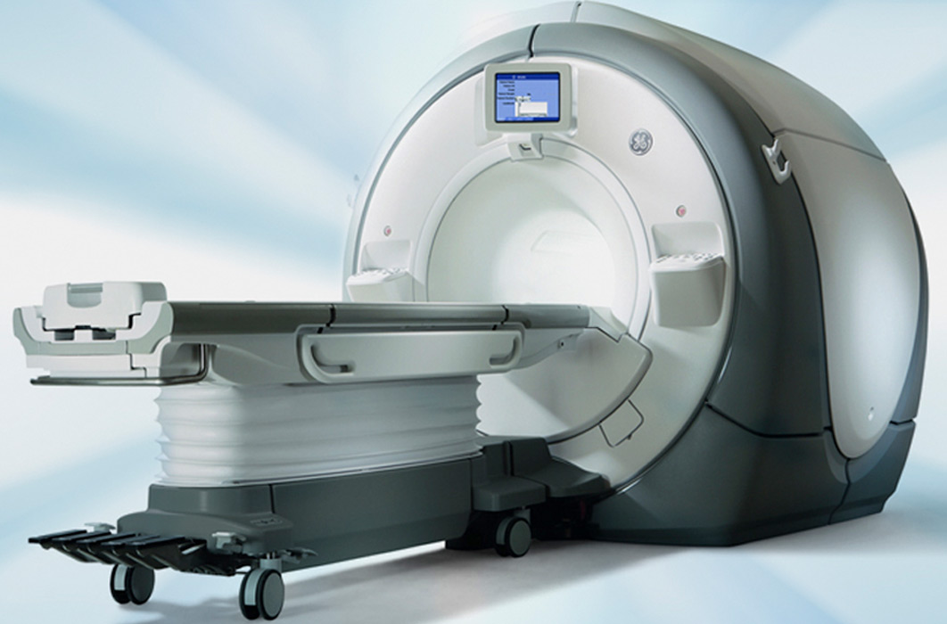
An MRI machine is a large cylindrical magnet. When you enter the MRI room, you will be asked to lie down on a bed, and the machine will move you and the bed into position, depending on the type of exam needed. During an MRI, you will be asked to be still for the duration of your scan, typically 20-45 minutes. Movement can blur the images and produce a lower-quality scan. The MRI machine can be loud, and you will hear loud tapping or thumping during the exam. Earplugs or earphones will be provided to you, and you can communicate with the technician through a microphone in the machine.
When your examination is over, you may resume your normal daily activities unless otherwise instructed by your doctor. One of our board-certified radiologists will review the images and send a report to your doctor.
What if I’m claustrophobic?
An MRI is painless. However, some claustrophobic patients may experience a “closed in” feeling when placed in the magnet. If this is a concern, please let us know prior to your appointment. Conscious sedation may be available depending on your preferred location.
Additionally, we offer both Open and Upright MRI at some locations. Open MRI, which does not completely surround the patient as with conventional MRI scanners, may be used to accommodate claustrophobic, obese or pediatric patients. Upright MRI, which allows imaging of a patient in an upright position, may be used to accommodate those who are claustrophobic, obese, or have a particular injury, making traditional MRI imaging difficult. If you are interested in either our Open or Upright MRIs, let us know when scheduling your appointment.
What if I require contrast?
Depending on the part of the body being examined, a contrast material may be injected to enhance the visibility of certain tissues or blood vessels. The contrast does not typically cause symptoms, though sometimes patients complain of nausea, vomiting, headaches or itching. More complex side effects are incredibly rare.